Fluoroscopy is a dynamic imaging technique that uses continuous X-ray beams to create real-time images of the internal structures of pets during surgical procedures. This allows veterinarians to visualize the area of interest in detail, making adjustments as needed throughout the surgery.
Fluoroscopy is commonly used in veterinary surgery for:
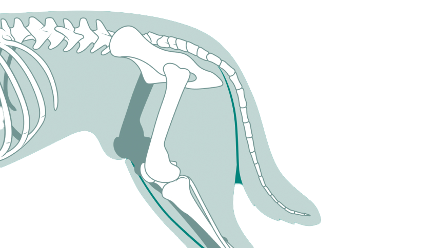
- Orthopedic Surgery: Assisting in the accurate placement of implants such as pins, plates, and screws.
- Fracture Repair: Ensuring proper alignment and stabilization of fractured bones.
- Joint Procedures: Visualizing joint structures and guiding the removal of loose bodies or other surgical interventions.
- Fractures: Real-time imaging ensures precise alignment and stabilization of bone fractures during repair.
- Joint Instability: Fluoroscopy helps in visualizing joint movements and guiding the placement of stabilization devices.
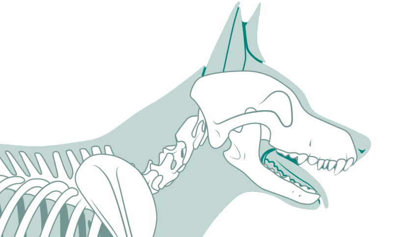
- Spinal Surgery: Providing detailed views of the spine during corrective surgeries for vertebral fractures or luxations.
Our areas of expertise
Advanced Imaging
Precision technology for accurate diagnosis and treatment planning.
Advanced Imaging
Back
Next
Transforming the way orthopedic care is delivered
For Vets
Refer your patients to our specialists using our streamlined online portal.
Referral portalFor Pets
Request a consultation or prepare for surgery with us and experience expert compassionate care every step of the way.
Request a consultation